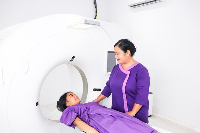
Contrast media for CT-scans are vital in angiography, enhancing blood vessel visibility and aiding radiologists in detecting anomalies. These agents, with ionic or non-ionic properties, improve tissue contrast, facilitating the assessment of vascular health by highlighting arteries, veins, and capillaries. Intravenous injection enables detailed visualization of vessel walls, blockages, and aneurysms, improving diagnostic accuracy and treatment planning. Safety has improved with modern low-osmolality agents and patient-centric practices, and future developments aim to enhance safety and imaging quality through biocompatible formulations and computational imaging techniques.
Contrast media play a pivotal role in angiography and vascular studies, enhancing diagnostic accuracy. This article delves into the world of contrast media, exploring their types, functions, and critical applications in CT scans. We examine how contrast enhancement amplifies visual detail, enabling radiologists to detect anomalies in blood vessels. Furthermore, we discuss safety considerations and emerging trends, including advancements in contrast agent technology tailored for CT scans. Understanding these aspects is crucial for maximizing the benefits of contrast media in modern medical imaging.
Understanding Contrast Media: Types and Functions
Contrast media play a pivotal role in angiography and vascular studies, enhancing the visibility and clarity of blood vessels and related structures on imaging tests like CT-scans. These agents are designed to improve the contrast between different tissues and organs, allowing radiologists to better detect anomalies or blockages within the vasculature.
There are various types of contrast media, each with unique properties and functions. For instance, ionic contrast media contain small molecules that easily move through blood vessels, providing rapid and distinct enhancement. Non-ionic contrast media, on the other hand, exhibit better compatibility with tissues, reducing potential allergic reactions. In CT-scans, contrast media are typically injected intravenously to highlight arteries, veins, and capillaries, enabling detailed assessment of vascular health and integrity.
CT Scans and Contrast Enhancement: A Powerful Combination
In angiography and vascular studies, the combination of CT scans and contrast enhancement offers a powerful diagnostic tool. Contrast media for CT-scan plays a pivotal role in enhancing the visibility of blood vessels and associated structures, allowing radiologists to gain detailed insights into their anatomy and any potential abnormalities. By injecting these specialized agents into the patient’s bloodstream, the vessels are highlighted, providing a clear contrast against surrounding tissues on the CT images.
This synergistic effect not only improves the accuracy of vascular assessments but also facilitates faster diagnosis and treatment planning. The use of contrast media in CT scans enables the detection of narrowings, obstructions, or abnormalities that might be missed in standard scans, making it an indispensable technique for comprehensive vascular evaluation.
The Role of Contrast Media in Vascular Studies
Contrast media play a pivotal role in vascular studies, enhancing the visibility and clarity of blood vessels and related structures during diagnostic procedures. In techniques such as angiography and CT-scan, these media are administered intravenously to improve image quality. By increasing contrast between the blood and surrounding tissues, healthcare providers can better visualize vessel walls, detect abnormalities, and assess blood flow patterns.
This is particularly crucial in identifying blockages, aneurysms, or other vascular conditions that might go unnoticed without the use of contrast media. In CT-scan, for instance, contrast agents allow radiologists to differentiate between various types of tissue and fluids, providing a more detailed and accurate picture of the vasculature. This, in turn, aids in making informed clinical decisions and guiding treatment strategies.
Safety Considerations and Future Directions
The safety profile of contrast media has evolved significantly over time, with strict regulations and guidelines in place to ensure minimal risks to patients undergoing angiography and vascular studies. While rare, potential adverse reactions include allergic responses, kidney damage, and cardiovascular events, particularly in patients with pre-existing conditions or compromised renal function. Therefore, careful patient selection, appropriate dosing, and close monitoring during procedures are essential. The choice of contrast media also plays a crucial role; modern, low-osmolality agents are generally safer and better tolerated than their older counterparts.
Looking ahead, future developments in contrast media for CT-scan promise to further enhance safety and imaging quality. Researchers are exploring biocompatible, bioabsorbable alternatives that could reduce potential long-term side effects. Additionally, advancements in computational imaging techniques may enable the optimization of contrast agent use, minimizing exposure while maximizing diagnostic yield. These innovations hold the key to improving patient outcomes and expanding the scope of vascular imaging studies.
Contrast media play a pivotal role in enhancing angiography and vascular studies, particularly in CT scans. By improving image quality, these substances enable precise diagnosis and effective treatment planning. As technology advances, ongoing research into safer and more effective contrast media for CT-scan applications holds the promise of better patient outcomes and enhanced clinical decision-making. Safety considerations remain paramount, but the benefits derived from contrast media continue to make them indispensable tools in modern radiology.
